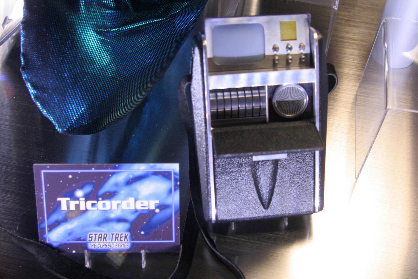
When Captain Kirk stepped out with a tricorder in hand in the very first episode of Star Trek in 1966, the data-sensing, -scanning and -analyzing gadget seemed to be a rather useful but very far-in-the-future piece of technology.
Nearly 60 years later, and the search to replicate a similar handheld diagnostic tool has seen scientists from Canada and Mexico develop the Swift Ray 1, which links to a smartphone and uses heat signatures and bacterial fluorescence to identify infected wounds.
Said to be the world’s first hyperspectral device so compact it fits in your pocket, the Swift Ray 1 can capture heat produced by an area of injury, which helps clinicians tell the difference between inflammation and infection.
In a clinical study of 66 wounds, the device deemed 20 non-inflamed, 26 inflamed and 20 infected. When subjected to a machine learning algorithm known as k-nearest neighbor the researchers found the gadget identified 100% of infected wounds and 91% of non-infected.
Because a healing wound will have a degree of redness to it, it can be notoriously difficult for a doctor to tell when a wound has gone from normal inflammation to potentially serious infection.
“Wound care is one of today’s most expensive and overlooked threats to patients and our overall healthcare system,” said corresponding author Robert Fraser of Western University and Swift Medical Inc. “Clinicians need better tools and data to best serve their patients who are unnecessarily suffering.”
This device attaches to a smartphone and links to an associated app. As well as medical-grade photographs, it also records infrared thermography images, which measure heat, and bacterial fluorescence imaging, which makes bacteria glow when exposed to ultraviolet light.
“Research has demonstrated bacterial imaging helps guide clinicians’ work to remove nonviable tissue, yet it cannot identify infection by itself,” explained first author Dr Jose Ramirez-GarciaLuna of McGill University Health Centre. “Thermography provides insight into the inflammatory and circulatory changes happening under the skin.”
While these sorts of visual diagnostic tools have been in development for some time, they generally specialize in one aspect, such as infrared thermography. Heat changes can be the result of inflammation, not infection, which is where bacterial fluorescence imaging comes into play.
While the researchers say that the results may still need further investigation – for example, a wound might be cool so deemed non-inflamed, but have a limited blood supply to the site of injury so instead has compromised healing.
However, an inexpensive, single multipurpose gadget like this could help clinicians intervene sooner, which is crucial for getting infections under control. And it’s also a little more streamlined than Kirk’s device …

The Swift Ray 1 is also able to ID across different skin pigments, which can be difficult for clinicians when based on eyesight triaging alone.
“This was a pilot study and follow up studies are planned,” cautioned Fraser. “In the future, patient populations with more wound types are required to validate across populations.”
The study was published in the journal Frontiers in Medicine.
Source: Swift Medical via Scimex
Source of Article