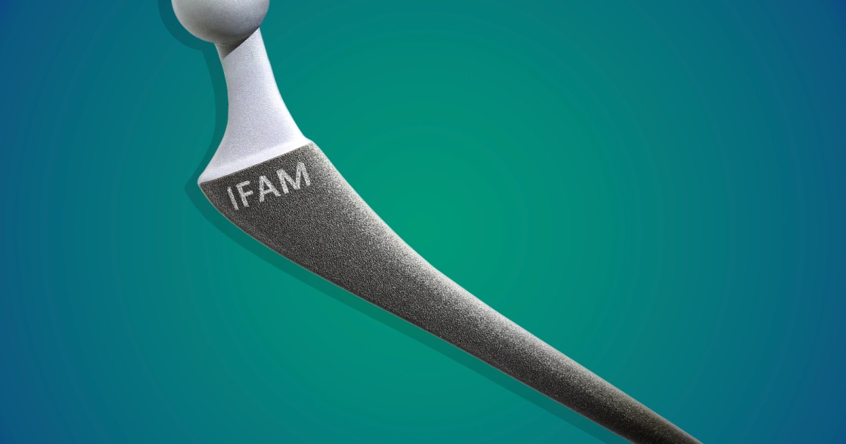
When a patient receives a titanium artificial hip, there’s always the risk of an infection developing at the interface between the metal and the bone. A new implant-coating process, however, is intended to greatly reduce that risk.
The technology was developed via a collaboration between Germany’s Fraunhofer Institute for Manufacturing Technology and Advanced Materials, and the Charité-Universtitätsmedizin Berlin. It’s part of the larger AntiSelectInfekt project.
The first step involves utilizing a laser to finely structure the surface of an existing titanium hip. Doing so leaves the surface of the metal full of microscopic pores, each one similar in shape to a tiny amphora – this means they’re wider at the bottom than they are at the top.
Next, a technique known as physical vapor deposition is used to apply a thin layer of silver to the metal. The silver, which has antimicrobial properties, coats the inside walls of each pore without actually filling it up.
Finally, right before implantation, the titanium hip is dipped in an antibiotic solution. That liquid is drawn into the pores.
Once the hip has been implanted, the antibiotic (which is tailored to the specific needs of each patient) starts flowing from the pores into the surrounding tissue. This helps keep any infections from developing immediately after surgery. The silver, however, releases bacteria-killing ions for the next several weeks, providing protection against infections throughout the healing phase.
As an added bonus, because the surface of the implant has been made more textured and porous, it’s better able to integrate with the adjacent bone. In fact, bone cells actually grow into the pores, helping anchor the implant to the bone over time.
Preclinical trials have shown that the coating process is indeed effective at preventing infections. It could also be applied to other types of titanium joints, such as shoulders and knees. In recent years, other approaches to killing bacteria at such implant sites have included the use of time-release antibiotic beads and flakes of graphene.
Source: Fraunhofer
Source of Article