One of the ways physicians can gain some forewarning of impending heart failure is through the detection of excess fluid in the lungs, and MIT researchers have developed a new machine learning tool that could offer them a helping hand. The algorithm is able to detect severe cases of this condition with a high level of accuracy, and the researchers behind it are hopeful it could be adapted to assist with the management of other conditions, too.
The research was carried out at MIT’s Computer Science and Artificial Intelligence Lab (CSAIL) and fits in alongside a raft of other promising machine learning and artificial intelligence tools that are reshaping medical diagnosis. Through the power of modern computing, these algorithms are able to look over medical imaging data to spot subtle but critical changes in human conditions that clinicians are unable to see, opening up some exciting possibilities.
This could mean picking up missed cancer diagnoses through CT scans or detecting signs of Alzheimer’s years before they become visible to doctors. We’ve also looked at how using artificial intelligence to analyze electrocardiogram results could help doctors determine patients most at risk of heart failure by identifying left ventricular dysfunction, and this new research follows a similar pathway, although focuses on a different mechanism.
Doctors use X-ray images of the lungs to assess fluid build up in patients at risk of heart failure, with the severity of the condition, known as “pulmonary edema,” then determining the course of treatment. The trouble is that these assessments are often based on such subtle features that they can lead to inconsistent diagnoses and treatment plans.
To bring machine learning into the fold, the team trained its algorithms on more than 300,000 X-ray images and their corresponding reports written by radiologists. This involved developing certain linguistic rules to make sure the data was analyzed consistently across the many samples.
“Our model can turn both images and text into compact numerical abstractions from which an interpretation can be derived,” says co-lead author of the paper Geeticka Chauhan. “We trained it to minimize the difference between the representations of the X-ray images and the text of the radiology reports, using the reports to improve the image interpretation.”
MIT
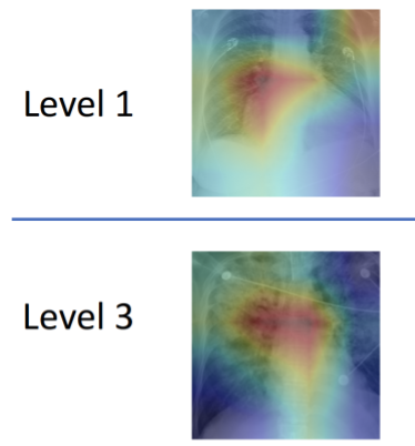
In putting it to the test, the team had the machine learning algorithm analyze single X-ray images and classify the severity of edema, ranging from 0 (healthy) to 3 (very, very bad). The algorithm was able to diagnose the correct level of edema more than half the time, but more impressively, was able to accurately diagnose level-3 cases 90 percent of the time.
The hope is that the tool can help doctors better manage heart issues, but edemas are linked to a range of conditions including sepsis and kidney failure, so the algorithm’s potential may be wide-ranging. The researchers are currently working to integrate the tool into the workflow of the emergency room at the Beth Israel Deaconess Medical Center in Boston in the next few months.
“This project is meant to augment doctors’ workflow by providing additional information that can be used to inform their diagnoses as well as enable retrospective analyses,” says PhD student Ruizhi Liao, who was the co-lead author of paper.
A research paper describing the technology can be accessed here.
Source: MIT
Source of Article