A team of UK scientists and engineers has demonstrated a novel technology for imaging the brains of infants and babies. The breakthrough is hoped to allow researchers new ways to investigate baby brain activity in natural environments without the need for expensive MRI machines.
“There is a lot we still don’t know about how the brain develops, and a big part of the problem is that studying the infant brain is really difficult with traditional scanners,” explains Rob Cooper lead on the project from University College London. “As any parent knows, 6-month old babies are very active; they move around all the time and are easily distracted. Using a technique like MRI, the subject has to remain completely still, which is almost impossible with babies unless they are asleep or sedated.”
The system presented in the study, published in the journal NeuroImage, is a new generation of wearable caps using high-density diffuse optical tomography technology (HD-DOT). The system is called LUMO, and the prototype tested came from Gowerlabs, a UCL spinoff company.
Each hexagonal tile on the cap contains three LED sources and four sensors. Near-infrared light is used to detect changes in brain oxygenation. Through these changes researchers can effectively map which parts of the brain are actively working in real-time.
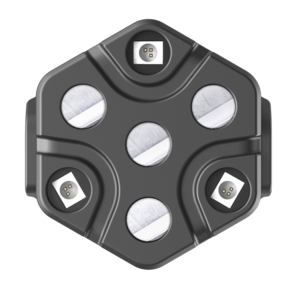
Gowerlabs
The closest imaging method to this new HD-DOT technology currently available to neuroscientists is called functional near-infrared spectroscopy (fNIRS). But fNIRS devices offer limited spatial resolution and can still require bulky headsets. Comparing the new HD-DOT devices to fNIRS measurements, the researchers say this new imaging technology is a dramatic improvement.
“Our results are consistent with prior low-density fNIRS measures based on similar paradigms, but demonstrate superior spatial localization, improved depth specificity, higher SNR [signal-to-noise ratio] and a dramatic improvement in the consistency of the responses across participants,” the researchers write in a new study. “Our data retention rates also demonstrate that this new generation of wearable technology is well tolerated by the infant population.”
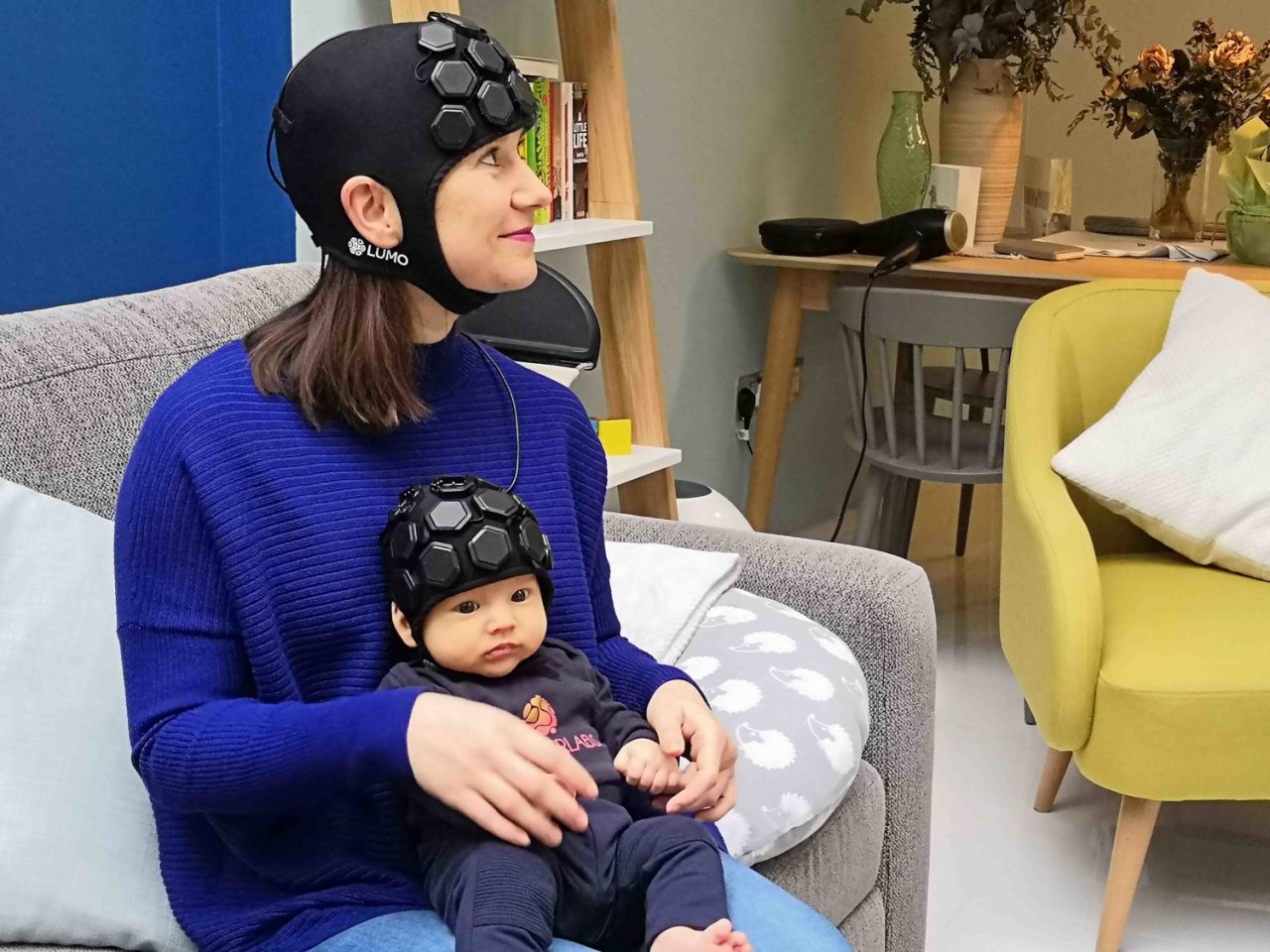
Gowerlabs
The HD-DOT system was tested on six-month old babies and found to be incredibly effective. Moving forward the researchers expect this new technology will allow neuroscientists extraordinarily novel insights into baby brain activity in natural environments.
“The approach we have demonstrated is safe, silent and wearable, and can produce images of brain function with better spatial resolution than any other comparable technology,” says Elisabetta Maria Frijia, one of the authors on the new study. “Our hope is that this new generation of technologies will allow researchers from a whole range of fields to learn more about how the healthy infant brain develops and establish new ways of diagnosing, monitoring and ultimately treating neurological conditions like autism and cerebral palsy.”
The new study was published in the journal NeuroImage.
Source: UCL
Source of Article