Currently, the best treatments for type 2 diabetes involve managing blood sugar levels, but recent research suggests that the brain may be a new drug target for longer-term control. Now, a pair of studies on rats and mice has investigated how a certain peptide works to induce remission for animals with diabetes, which could lead to new breakthroughs in human treatments.
Previous studies have found that just one injection of a protein called fibroblast growth factor 1 (FGF1) into the brains of mice with type 2 diabetes was enough to return them to regular blood sugar levels for weeks or even months. But the mechanism behind it remained mostly unknown. Now, two related studies have investigated how it works.
The first study charted the changes of gene expression in a variety of brain cell types after FGF1 injections, while the second looked into how other brain structures help to sustain diabetic remission. Both studies focused on the hypothalamus, a small region of the brain that regulates hunger, food intake, energy use and, importantly, blood sugar levels.
In the first study, the team surgically injected FGF1 into the brains of mice with type 2 diabetes, then monitored the changes in gene expression from different brain cell types. They found that the structural glial cells responded more strongly than neurons did.
But the key finding was that FGF1 acted on glial cells to strengthen the melanocortin signaling system. This brain circuit controls feeding, body weight and blood sugar, and reduced activity in this system has been linked to diabetes development in humans and rodents. Indeed, the team found that blocking melancortin signaling prevented diabetes remission, even after FGF1 injection.
The team says that this crucial finding suggests that glial cells and the melancortin signaling system could be new drug targets for diabetes.
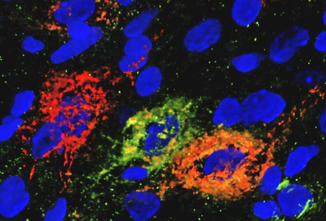
Kim Alonge/Schwartz Lab
The second study focused on structures called perineuronal nets, which surround and support neurons and their connections. The team found that rats with type 2 diabetes have fewer perineuronal nets in their hypothalamus than healthy animals.
Interestingly, the injection of FGF1 quickly reversed this loss, with new nets growing in the hypothalamus of treated rats. To test the inverse, the researchers removed nets from the brains of the animals and found that FGF1 had a harder time managing diabetes.
The team speculates that perineuronal nets might be helping to strengthen the activity of neurons that control melanocortin signaling. Both studies point to new avenues for future research to investigate, in the hopes of eventually developing longer-term treatments for diabetes management.
“Until recently, the brain’s ability to normalize elevated blood sugar levels in diabetic animals was unrecognized,” says Dr. Michael Schwartz, an author on both studies. “Our international teams’ latest findings chart a path towards a more complete understanding how this effect is achieved. These insights may one day inform therapeutic strategies for inducing sustained diabetes remission, rather than simply lowering blood sugar levels on a day-to-day basis as current treatments do.”
The first study was published in the journal Nature Communications, while the second appeared in Nature Metabolism.
Source: University of Washington
Source of Article